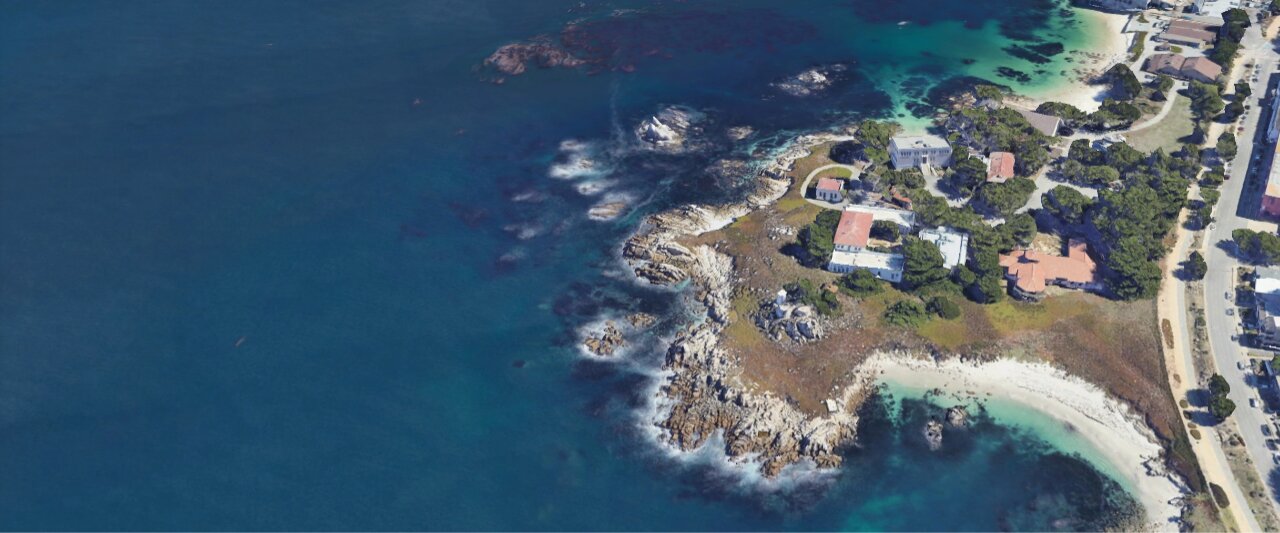
The Barone Laboratory is located at the
Hopkins Marine Station and is part of the Biology Department of  Stanford University.
We explore how variation in cell behaviours that determine the physical properties of tissues contribute to the evolution of development.
Marine embryos help us in our research: meet the sea star and sea urchin embryos
Although early development of sea star and sea urchin is similar in many ways, there are key differences in their cell and embryonic shapes. For instance, sea urchin embryos are more compact than sea stars' and cell size is more homogenous in the sea star than in the sea urchin. We ask how these differences in embryonic shape affect patterning and cell fate decisions.
Our method of choice is live imaging. It allows us to carefully observe the shape of the whole embryo and the behaviours of individual cells within it.
We combine molecular biology, cell biology and biophysics approaches to understand how the physical properties of cells determine embryonic shapes and their variation. Below you will see an example of how we use micropipette aspiration to measure the surface tension of sea star cells.